Introduction: Why Animal Tissues Trip Up NEET Aspirants
NEET comes with at least 3 or 4 questions directly from animal tissues every year, and most students lose those marks not due to lack of study but due to memorising without understanding. On the surface, it seems simple – four types of animal tissues and that’s it. But in the real exam, they test you on subtypes, structure, which tissue lines which organ, etc., and why.
A study of the human body reveals the presence of organs such as a heart, a stomach and a lung. What are those organs really composed of? So the answer is tissues. Prior to organs, there are tissues. There are cells before the tissues. There it all starts. Here are the things that make this topic really difficult. If read quickly, all four animal tissues look alike. Both epithelial and connective tissue “guard” the body but in different ways. Smooth muscle and cardiac muscle are both involuntary, but they certainly have very different structures.
This blog is a single-stop guide for you to learn animal tissues class 11 in a way that is entirely beneficial for your NEET 2026 and Board exams. We will discuss Tissues, their classification, structure-function-location of all four types of tissues, their subtypes, real NEET PYQs with explanation and FAQs. No fluff. Only what is being tested, exactly.
Read More:
- Important Biology Diagrams for NEET 2026: Biology Diagrams for Botany & Zoology
- Connective Tissue in Biology for NEET: Types, Functions & Notes
What Is a Tissue? The Definition You Should Actually Remember
A tissue is a group of similar cells, often held together by an extracellular matrix, that arises from a similar origin and works together to perform a specific function. But in some cases, certain complex tissues (like the blood or vascular tissue in plants) contain visibly different cell types working together as a functional unit.
It was the French anatomist Xavier Bichat who coined the term ’tissue’ in 1801. He is described as the ‘Father of Histology’, and the term histology refers to the study of tissues at a microscopic level. A fact NEET has tested before so remember this.
Quick fact: An organ is formed by the combination of different tissues to carry out a similar function. Organ systems are formed when organs work together. In turn, tissues are the building blocks of anything above them.
Check Out: Difference Between Plant Cell and Animal Cell in Biology: Notes, Table & Diagram for NEET
What Are Animal Tissues? Origin and Classification
As per NCERT, animal tissues are defined as: “Tissues found in multicellular animals that originate from the three primary germ layers – ectoderm, mesoderm, and endoderm, formed during embryonic development.”
The four germ layers are the source of all the body tissues and organs. The difference between animal tissue cells and plant tissue cells is simple and fundamental: Plant cells have a cell wall, while animal cells lack one. However, they are bound together by intercellular junctions and intercellular cementing substances.
This is one of the most commonly asked points in the difference between plant tissue and animal tissue:
| Feature | Plant Tissue | Animal Tissue |
| Cell wall | Present | Absent |
| Living at maturity | Can be dead (e.g., sclerenchyma) | Mostly alive |
| Types | Meristematic & Permanent | Epithelial, Connective, Muscular, Nervous |
| Origin | Single layer (protoderm, etc.) | Ectoderm, Mesoderm, Endoderm |
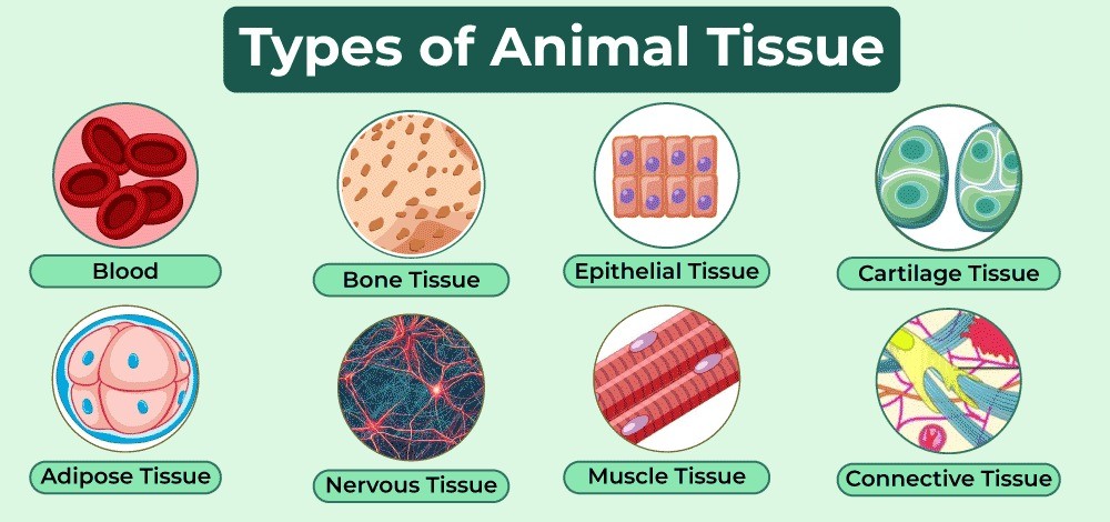
Based on their location and function, animal tissues are classified into four types:
| Tissue Type | Origin | Primary Function | Location (Examples) |
| Epithelial Tissue | Ectoderm, Endoderm, Mesoderm | Protection, secretion, absorption | Skin, gut lining, lungs |
| Connective Tissue | Mesoderm | Support, binding, transport, storage | Bone, blood, tendons |
| Muscular Tissue | Mesoderm | Movement and contraction | Heart, skeleton, hollow organs |
| Nervous Tissue | Ectoderm | Signal conduction, coordination | Brain, spinal cord, nerves |
Check Out: Most Scoring Units & Chapters in Biology That Cover 60% of NEET 2026 Biology Paper
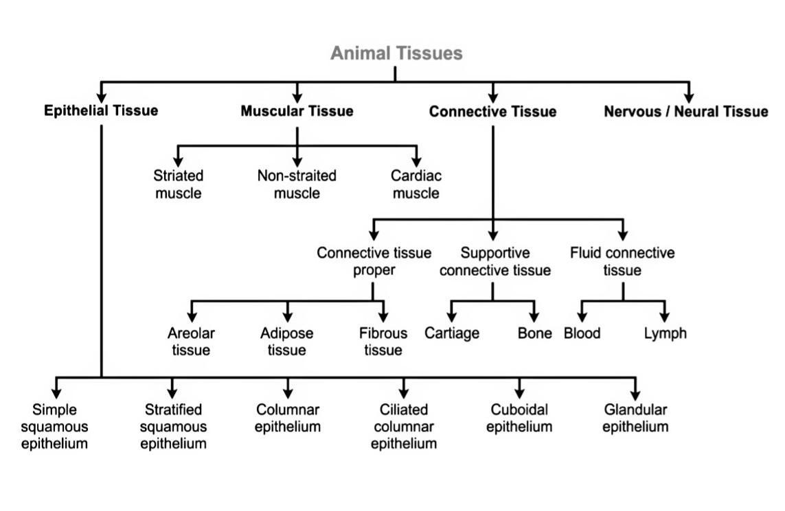
4 Types of Animal Tissues: Structure, Function, Location & Subtypes
1. Epithelial Tissue – The Body’s Covering and Lining
Epithelial tissue is like wallpaper; it lines all surfaces, all cavities, all hollow organs. Anything that comes in contact with the external environment or internal body fluids is covered by epithelial tissue.
- Structure: Cells are densely packed and have little intercellular space. No direct blood supply (avascular) and the tissue lies on a basement membrane, separating it from deeper tissues.
- Function: Protection (from physical damage, pathogens, and drying out), secretion of enzymes and hormones, absorption of nutrients, excretion of waste, and filtration of substances.
- Location: Outer layer of skin, inner lining of the digestive tract, inner walls of blood vessels, kidney tubules, lung alveoli, and ducts of glands.
Exam Insight: Epithelial cells are avascular (no blood vessels), so they absorb nutrients from the connective tissue below by diffusion. That’s why the basement membrane is so significant, as it serves as the link between the two.
Subtypes of Epithelial Tissue
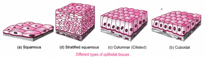
There are two broad categories based on layering: Simple Epithelium (single layer) and Compound Epithelium (multiple layers). This is a classic NEET question on simple epithelium vs compound epithelium.
A) Simple Epithelium
A single cell layer present on the basement membrane. It is only one cell thick and therefore good for absorption, secretion and diffusion.
- Squamous Epithelium: These are very flat, scale-like cells that fit together like floor tiles. The cells look irregular and polygonal when viewed from above. Found in the walls of blood capillaries, lung alveoli, and Bowman’s capsule of the kidneys. Function: diffusion and filtration.
- Cuboidal Epithelium: Cells are roughly cube-shaped, with a round nucleus in the centre. Found in the kidney tubules, salivary gland ducts, and thyroid follicles. Function: secretion and absorption.
- Columnar Epithelium: Found lining the stomach, intestine, and gall bladder. They carry out the secretion of mucus and digestive enzymes, and the absorption of digested food.
- Ciliated Epithelium: A columnar cell is any cell that is shaped like a column and has cilia, which are tiny, hair-like, motile projections on the free surface. Found in the Fallopian tubes and are present in the respiratory tract (trachea and bronchi). Use: to clean out mucus, dust and unwanted material.
- Glandular Epithelium: Specialised cells that secrete substances. They produce and release substances like mucus, hormones, enzymes, or sweat. Goblet cells are a classic example: unicellular mucus-secreting glands found within columnar epithelium.
B) Compound Epithelium
Two or more cell layers. The primary function is protection; it is not well-suited for absorption because the multiple layers slow down transport.
- Stratified Squamous Epithelium: Multiple layers of flat cells. The outermost layers are dead and filled with a protein called keratin. Found in the skin, oral cavity, oesophagus. Protects against mechanical wear and tear.
- Transitional Epithelium: A unique type found only in the urinary bladder and ureter. Can stretch and relax as the bladder fills and empties. This is why it is called transitional; it transitions between shapes.
Check Out: Difference Between Biotic and Abiotic Factors in Ecosystem
2. Connective Tissue – The Body’s Framework
Connective tissue is the most abundant and widely distributed tissue in the body. What makes it unique among all types of animal tissues is its extracellular matrix, a non-living substance surrounding the cells that can be fluid (like in blood), semi-solid (like in cartilage), or solid (like in bone).
- Structure: Cells scattered in a matrix. The matrix contains fibres mainly collagen fibres (which give strength) and elastin fibres (which give flexibility). The cells responsible for secreting these fibres are called fibroblasts, found mainly in areolar tissue.
- Function: To support and shape organs, to hold the various tissues and organs together, to store fat for energy, to defend the body (by means of immune cells), to transport materials (blood) and to repair damaged tissue.
- Location: Tendons, ligaments, cartilage, bone, blood, adipose tissue, dermis of skin, and the connective tissue sheets (fascia) around muscles.
NEET Alert: NEET 2024 re-examination had a question that asked which type of connective tissues secrete collagen and elastin fibres. The answer is fibroblasts found in areolar tissue. Be careful not to mistake fibroblasts for osteoblasts (bone cells) or chondroblasts (cartilage cells).
Subtypes of Connective Tissue
A) Loose Connective Tissue
A loose arrangement of cells in a semi-fluid matrix.
- Areolar Tissue: This is the most common and prevalent connective tissue. Located inside the skin, between muscle and muscle, surround blood vessels. Has fibroblasts, mast cells and macrophages. The main function of areolar tissue is to support, cushion and allow movement of fluids between tissues. It is also involved in repair and immune functions.
- Adipose Tissue: Found under the skin (subcutaneous fat), around the kidneys, and behind the eyeballs. Functions: energy storage, insulation against cold, padding and protection of organs, and hormone secretion (e.g., leptin).
B) Dense Connective Tissue
Contains closely packed collagen fibres with fewer cells, mainly fibroblasts. Much stronger than loose connective tissue.
- Dense Regular: All collagen fibres run in one direction, forming strong parallel bundles. Found in tendons (muscle to bone) and ligaments (bone to bone). Ligaments are dense, regular connective tissue, a fact NEET 2023 tested directly.
- Dense Irregular: The fibres are arranged in various directions, so that the tissue is strong against forces acting from different directions. Present in the dermis of skin and in a capsule around organs such as the kidney.
C) Specialised Connective Tissue
These have highly modified matrices suited for specific functions.
- Cartilage: Here, the matrix is semi-solid and rubbery and composed of a protein called chondrin. The cartilage is avascular, meaning that there are no blood vessels, and the cells of the cartilage receive nutrients by diffusion through the matrix; after damage, cartilage heals slowly. Located in joints, outside of ear, nose tip and tracheal rings. Supports the structure and provides smooth surfaces.
- Bone: Hard matrix with calcium phosphate and calcium carbonate. Contains osteocytes in spaces called lacunae, arranged in rings called Haversian systems around a central canal. Provides a rigid framework and protects organs (e.g., ribs protect the lungs) and serves as a reservoir of calcium and phosphorus.
- Blood: The most unique connective tissue, its matrix is liquid, called plasma. Plasma is about 55% of blood and carries dissolved proteins, nutrients, hormones, and waste. The cells suspended in plasma include RBCs (carry oxygen via haemoglobin), WBCs (immune defence), and platelets (clotting). Blood is the only fluid connective tissue, and it transports everything the body needs to distribute or remove.
3. Muscular Tissue – The Tissue That Moves You
Muscular tissue is made of elongated cells called muscle fibres that can contract and relax. This ability to contract is because of two protein filaments inside the cells, actin and myosin. Every movement your body makes, blinking, breathing, and pumping blood, is muscular tissue at work.
- Structure: Long cells (called muscle fibres or myocytes) that contain multiple contractile units. Rich in mitochondria to supply the large amount of energy needed for contraction.
- Function: Contraction leading to movement of bones, movement of substances through hollow organs, and continuous pumping of the heart.
Subtypes of Muscular Tissue
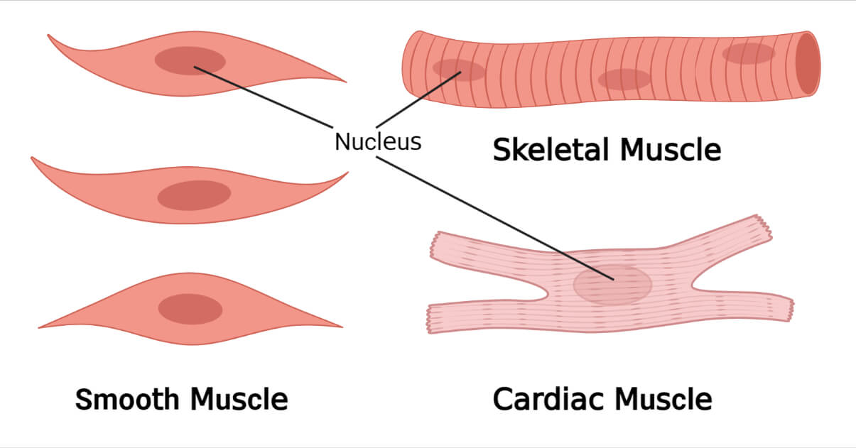
- Skeletal (Striated) Muscle: Attached to bones through tendons. Has alternating dark (A-band) and light (I-band) bands visible under a microscope, called striated. They are under voluntary control; you consciously decide when to use them. Example: biceps, triceps, quadriceps. They contract fast and powerfully but fatigue quickly.
- Smooth (Non-striated / Visceral) Muscle: Present in the walls of hollow organs, such as the intestine, stomach, uterus, urinary bladder, and blood vessels. Involuntary control. Composed of cells that are spindle-shaped and have a single nucleus. No striations. These are under involuntary control: you don’t consciously tell your intestines to move. Their contraction rate is slow and steady and they cause the involuntary actions such as peristalsis (food being pushed through the gut).
- Cardiac Muscle: Found only in the heart wall (myocardium). Like skeletal muscle, it is striated it shows light and dark bands. But unlike skeletal muscle, it is involuntary; your heart beats automatically without conscious effort. Has branched cells connected by intercalated discs, which allow rapid signal transmission, so the whole heart contracts as one unit.
NEET Favourite: Cardiac muscle is the only muscle that is both striated AND involuntary. Skeletal is striated and voluntary. Smooth is non-striated and involuntary. This comparison is tested almost every year.
4. Nervous Tissue – The Body’s Communication Network
The master coordinator of the body is nervous tissue. It gathers information from the environment (with the sensory organs), sends information to the brain and spinal cord for processing and sends out commands (to muscles and glands). It develops into the brain, spinal cord, and all peripheral nerves. It is formed from the ectoderm of the embryo – the same germ layer that gives rise to the skin.
- Structure: Nervous tissue is made of two main components: neurons (the signal-transmitting cells) and neuroglia (the supporting cells).
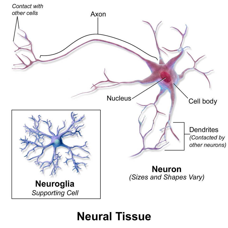
Neurons (Nerve Cells)
These are the basic structural and functional units of the nervous system. A typical neuron has three parts:
- Cell body (Cyton): Contains the nucleus and most of the metabolic machinery. This is where the neuron’s maintenance happens.
- Dendrites: Short, branched projections from the cell body that receive signals from other neurons or sensory receptors.
- Axon: A single, long projection carrying electrical signals from the cell body to the next neuron or to an effector (such as a muscle). The axon is usually coated with a fatty substance known as myelin sheath that acts as insulation to an electric wire, allowing the signal to conduct much faster. The end of the axon contains synaptic knobs that send out neurotransmitters to communicate with the next cell.
Types of Neurons (based on function):
- Sensory (Afferent) Neurons: Carry signals from sensory organs (eyes, skin, ears) towards the brain or spinal cord.
- Motor (Efferent) Neurons: Carry signals away from the brain/spinal cord to muscles and glands, triggering a response.
- Interneurons (Association Neurons): Located only in the brain and spinal cord. They link sensory and motor neurons and are important in thinking, memory, and reflexes.
Neuroglia (Glial Cells)
These are non-neuronal cells of the nervous tissue. They do not transmit nerve impulses, and are essential for the health and function of neurons. They nourish neurons by providing nutrients, protect them by forming the blood-brain barrier, produce myelin (Schwann cells in peripheral nerves), and remove dead cells and debris. There are about 10 times more glial cells than neurons in the nervous system.
- Function: The reception of stimuli (by sensory receptors), conduction of nerve impulses (along neurons) and coordination of all the activities of the body.
- Location: Brain, spinal cord and all peripheral nerves in the body.
Check Out: NEET Previous Year Question Papers with Solutions – Free PDF Download
Animal Tissue NEET PYQs: Important Questions from Past Papers
These are real questions from past NEET and AIPMT papers on animal tissue NEET questions and animal tissue PYQ NEET pattern:
- NEET 2024 (Re-exam): “In which of the following connective tissues do cells secrete fibres of collagen or elastin?” Options: cartilage, bone, adipose, blood, and areolar tissue. Answer: Areolar tissue because it contains fibroblasts.
- NEET 2023: “Statement I: Ligaments are dense, irregular tissue. Statement II: Cartilage is dense, regular tissue.” Both statements are incorrect. Ligaments are dense, regular, and cartilage is specialised connective tissue.
- NEET 2022: “Gap junctions allow ions to pass quickly between cardiac muscle cells for synchronised contraction.” True, these gap junctions are part of intercalated discs.
- AIPMT (Classic): In the walls of capillaries, what kind of epithelial tissue would you observe? Answer: simple squamous epithelium because it is thin enough to allow gases and nutrients to pass through easily.
- NEET 2019: “Which is a correct match between tissue type and its characteristics?” Tests knowledge of matrix composition in bone vs cartilage.
Pattern insight for structural organisation in animals class 11: NEET loves to ask about the subtypes of connective tissues and characteristics of cardiac muscle tissue more than anything in this chapter. If you have less time, then preparing those two areas can offer maximum return on your study time.
Frequently Asked Questions on Animal Tissues
1. What are the 4 types of animal tissues?
The four types of animal tissues are epithelial tissue, connective tissue, muscular tissue, and nervous tissue. Each is classified based on its location and function in the body.
2. What is the function of connective tissue in animals?
The functions of connective tissue are to support structure, to join organs and tissues together, to store fat for energy, and to transport substances. Blood is also a connective tissue, the only fluid tissue and it carries oxygen, nutrients and waste around.
3. What is the difference between plant tissue and animal tissue?
The principal difference between plant tissues and animal tissues is the presence of a cell wall in plant cells, which is absent in animal cells. At maturity, plant tissues can be dead (e.g., sclerenchyma) while most animal tissues are alive. The tissues of animals fall into four main groups (epithelial tissues, connective tissues, muscular tissues, and nervous tissues), and in plant tissues can be divided into two types, meristematic and permanent tissues.
4. Which animal tissue is responsible for movement?
Animals move by the action of the muscular tissues. Skeletal muscle is a voluntary muscle that moves the skeleton. Smooth muscle transports substances in hollow organs. The cardiac muscle is the muscle responsible for the continuous pumping action of the heart, without any conscious effort.
5. What is histology, and who is the father of histology?
Histology is the microscopic study of tissues, their structure, composition, and function. Xavier Bichat (1771–1802) is called the Father of Histology. He introduced the term ’tissue’ in 1801 and was the first to systematically classify body tissues, even without the use of a microscope.
Written By: Saumya Sarin (Content Writer at Motion Education)
Reviewed By: Senior Biology Faculty (Motion)
Last Updated: June, 2026


